
Physical Acoustics Laboratory
Department of Engineering Science
Research
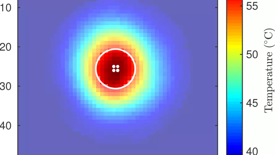
Infrared thermal imaging (FLIR) for monitoring heating due to high intensity focused ultrasound
Spatiotemporal surface temperature elevations are measured using an infrared camera (A655sc; FLIR Systems, Kent, UK) and used to calculate the spatially-dependent thermal dose delivered to ex-vivo tissues.
Collaborators
Image

Biomedical Ultrasonics, Biotherapy and Biopharmaeuticals Laboratory (BUBBL)
Institute of Biomedical Engineering, University of Oxford
Image

The Institute of Acoustics
School of Physics, Nanjing University
Image

Applied Optics Group
School of Physical Sciences, University of Kent